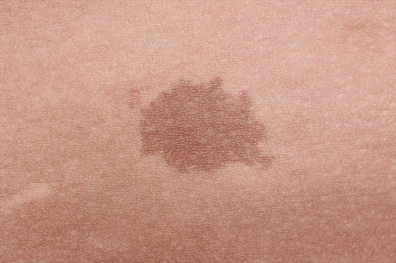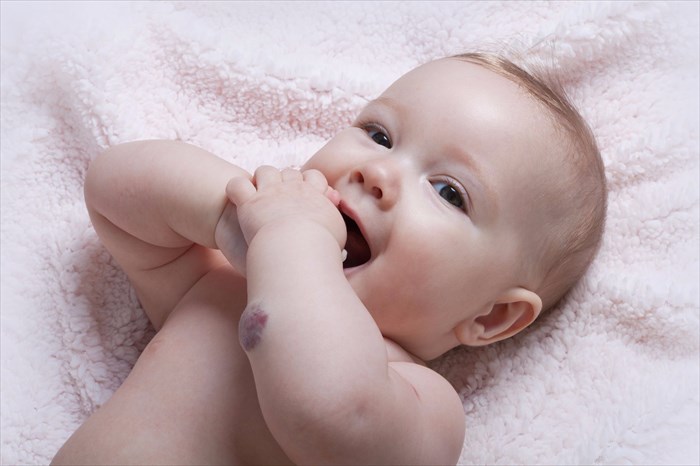
Vascular (i.e. birthmarks that result due to the abnormal growth or formation of blood vessels and vessel cells) and pigmented birthmarks (i.e. those that occur due to an overgrowth of the cells that produce colour in the skin) are categorised in relation to their composition.
Vascular birthmarks
Vascular birthmarks appear flat on the skin’s surface although in some instances, may be slightly raised. They may also change or evolve during the course of a person’s life. Colourations range from a light pink to a dark purple, and the markings vary in size. Certain characteristics are specific to the different subtypes of these birthmarks. Colloquially, these are referred to as ‘red birthmarks’4.
Typically, vascular birthmarks develop before birth and appear shortly after delivery. Some may initially appear rash-like and become more noticeable when an infant experiences body temperature changes or cries. These marks may itch, and possibly form open sores / ulcers and sometimes bleed.
These birthmarks are associated with the malformation of capillaries (blood vessels) and/or vascular structures. Medical professionals also classify vascular birthmarks as either ‘tumours’ or ‘malformations’. This is because the clinical course of these formations differs, and thus so do treatment recommendations and the overall outlook.
A vascular tumour is effectively a ‘benign blood vessel mass’ which is composed of actively multiplying blood vessels. Vascular malformations, on the other hand, contain dilated blood vessels which are not actively multiplying. Tumours grow rapidly, while malformations dilate gradually. Malformation may not shrink, while tumours can.
Some of the most common forms of vascular birthmarks include:
Haemangioma (or infantile haemangioma)
These birthmarks (pronounced he-man-gee-oh-ma), are classified as vascular tumours5 and are typically characterised as red and raised ‘strawberry’ markings (pink, red and sometimes bluish in colour) which may initially present as small, flat blemishes, typically on a large area of the face or body (trunk).
They can increase in size, a unique characteristic of these birthmarks. Growth rate is often quickest during a baby’s first few months of life (and up to 12 months)6. While further growth may occur beyond that, it is considerably slower, if at all. Generally, these marks remain at a fixed size for a period but may then reduce (in a process of involution and regression). Certain markings can fade somewhat over time becoming almost white-grey in colour and flattening out, while others may stretch and deform (causing skin puckering), particularly if the marking is large.
Interestingly, these types of birthmarks appear to be most common in twins, females and those with lighter skin tones. They are also associated with premature and low birth weight babies. The cause of this is not yet known.
The location of a haemangioma can occasionally be medically significant and lead to potential health related complications. Such locations include those near the eyes (troubling vision capacity), mouth (leading to feeding difficulties), in the throat area (influencing breathing function), or anywhere else on the body that becomes an open sore or ulcer (and occasionally bleeds).
A doctor cannot predict how big a birthmark will grow or by how much it may fade either. Although a very obvious marking to have, unless there are multiple growth markings (in which case a doctor may wish to medically determine the possibility of internal haemangiomas) or the birthmark causes some functional difficulty due to its location, these are generally left alone. Treatment may however be considered for cosmetic reasons if markings are large and occur on the scalp, face or neck, and cause the affected person emotional distress due to the influence on appearance.
Haemangiomas fall into two classifications:
- Superficial (or capillary) haemangiomas (strawberry mark / haemangioma, nevus vascularis, capillary haemangioma, haemangioma simplex)
- Deep (or cavernous) haemangiomas (cavernoma, cavernous haemangioma angioma cavernosum).
Superficial variations tend to be redder in colour (due to closely packed blood vessels) and raised as the affected abnormal blood vessels are close to the surface of the skin. Common areas in which they occur include the face, scalp, chest and back.
Deep haemangiomas have a more bluish tinge by contrast, as the affected blood vessels are located in the deeper skin layers. These markings are not always present at birth and may only appear within the first few weeks of a baby’s life. They can be characterised as a red-blue spongy tissue mass / bulge (sometimes filled with blood). Some may even disappear on their own during childhood.
Haemangiomas which involve both the superficial skin layers and deeper tissues beneath are known as compound haemangiomas.
It is very unusual (rare) for a baby to develop more than one haemangioma – however, if this does occur and multiples are present (especially if 5 or more have formed), the possibility of internal variations will be considered by a medical doctor and the baby will be evaluated accordingly.
A doctor will likely recommend a liver ultrasound, complete blood count (blood test) and liver function test. Tests are recommended to determine the depth of a haemangioma (which can be seen) and whether it is hindering the normal function of the internal organs in any way. Although rare, some internal haemangiomas can develop in the body’s organs, like the liver, kidneys, lungs and brain, as well as the in the airways and gastrointestinal tract, and thus do not always show up as external lesions.
Macular stains / telangiectatic nevus / nevus simplex
These vascular malformation markings are often called ‘stork bites’ (or marks) and ‘salmon patches’, and present as patches of slightly reddened or pink skin7. An accumulation of capillary blood vessels is generally believed to influence the formation of these markings.
Salmon patches which form on the facial area are often referred to as an ‘angel kiss’. They can also occur on the eyelids, forehead or in the space between the eyes, as well as the upper lip or tip of the nose. Stork bites are so called because of their most common location, the back of the neck (just above the hairline) where the proverbial ‘stork’ would have carried the newborn in its beak.
Angel kisses most often fade over time (sometimes even by age two), while stork bites generally don’t but can be covered somewhat by hair growth. Most of these birthmarks will never require any kind of medical intervention or treatment.
Port wine stains / nevus flammeus
These low-flow vascular malformation birthmarks are normally present at birth causing red or purplish markings somewhere on the body. Common in the facial area, it is believed that port wine stains are linked to capillary malformation and depending on the extent, range in size. Markings may darken as the child grows, and the affected skin may thicken or even develop bumps or ridges, giving it an irregular, pebble-like texture.
These birthmarks can sometimes, although rarely, be linked to or coincide with medical problems, like Klippel-Trenaunay syndrome (a rare congenital vascular disease that presents with birthmarks, varicose veins, as well as excess bone and soft tissue growth)8 or Sturge-Weber syndrome (a neurological disease characterised by port wine birthmarks on the forehead, scalp or eyes, affecting blood vessels on the same side of the brain that the marking occurs on the face). Both conditions require regular evaluation. Also rare but possible, is an association with the development of glaucoma and seizures if the marking is on the forehead, sides of the face or around the eyes.
If these types of birthmarks are merely a cause of emotional distress, laser therapy treatment can help to lessen the appearance of these types of markings.
Pigmented birthmarks
Pigmented birthmarks occur due to an excessive accumulation of pigment or melanin in the deeper layers of skin (known as dermal melanosis) which produces dark markings. Melanin accumulation which is contained in melanocytes (pigment cells) produces markings called nevus (nevi are typically small, but can also become fairly large). A considerable lack of melanin deposition, in contrast, can produce markings which are lighter than the remainder of the body’s skin.
These marks may be flat or appear slightly raised on the skin (sometimes with a wrinkled appearance), but can also change / increase in size as a baby grows. Colourations range from tan, to brown, blue, blue-grey and black (almost like bruises). Colouration changes can also occur during the course of life, and are influenced by hormone fluctuations, especially during the teenage years, or sun exposure, and as a result may develop itchiness and an increased tendency to bleed.
Some of the most common forms of pigmented birthmarks include:
Mongolian spots (dermal melanocytosis / slate grey nevus)
These birthmarks are often likened to bruises9 because of their bluish colouration (these hues include blue-grey, blue-black or blue-brown). Mongolian spots commonly occur on the lower back, buttocks, trunk of the body or arms, and more frequently affect individuals with darker skin tones. These marks do tend to fade, often within the first 4 years of life (but aren’t likely to disappear completely), and are considered harmless (not requiring medical treatment).
Café-au-lait spots / Café-au-lait macules
These marks commonly occur at birth, but can also develop during the initial few years of a child’s life. Markings are usually a light tan or brown colour (almost like milky coffee, hence the name), and oval in shape. These birthmarks don’t typically fade with age, may even darken over time and remain for life. They can increase in size as a child grows.
One or two markings (pairs) are common but it is also possible to have more. If several markings occur (at least 6), that are larger than a coin, a doctor may wish to evaluate a child for a genetically inherited disorder associated with these birthmarks, known as neurofibromatosis type 1 (this condition is characterised by abnormal cell growth affecting nerve tissues and resulting in neurofibromas or tumours)10.
Congenital nevi (moles) / congenital melanocytic nevus
When present at birth, these markings commonly known as moles, are also considered birthmarks (those that develop later in life are not generally classified as such), and do carry a degree of risk for developing skin cancer. ‘The larger the mole, the greater the risk’ is the general rule of thumb. Moles can occur anywhere on the body, with many forming on the head, scalp or neck, as well as the trunk.
These markings are typically either light brown in colour or darker (almost black in darker skinned individuals). They may be flat, raised, lumpy, and irregular in shape. They can decrease in size as a baby grows, but can darken with age or exposure to sun and develop hairs as a child reaches puberty. The use of birth control pills (oral contraceptives) or pregnancy can also darken these marks.
A doctor will normally check these when they are noticed at birth and recommend further check-ups should any characteristic changes take place down the line.
What is the silvermark and is it a birthmark?
A naturally silver streak of hair, typically located at the left or right side of the head where the forehead and hairline meet, is called a silvermark. It is considered a birthmark, although it affects the hair more than the skin.
For many that way inclined, these silver streaks are regarded as a blessing, often admired rather than being a source of embarrassment or emotional distress. Some may call it by its more colloquial name – 'the witch’s streak' – because of the aesthetic look of a silver streak of hair in the front portion of the hairline, which for many has a mysterious and charismatic feel.
This type of birthmark is hereditary and has been known to occur in families with susceptible genetic structures. The mark generally appears in childhood with the colour growing out from the root along the entire length of hair.
The birthmark doesn’t really fall into the vascular or pigmented categories, but may have something to do with melanocytes that are responsible for producing melanin pigment (which influences hair colour) and proteins in the hair. If these proteins shut down (genes do not mutate) and an abnormality occurs (i.e. protein is not produced), a portion of affected hair is devoid of pigment-making structures and grows without pigment (colour), producing a streak.
References:
4. MedlinePlus. August 2018. Birthmarks - Red: https://medlineplus.gov/ency/article/001440.htm [Accessed 29.08.2018]
5. US National Library of Medicine - National Institutes of Health. May 2014. Vascular Tumours: https://www.ncbi.nlm.nih.gov/pmc/articles/PMC4078200/ [Accessed 29.08.2018]
6. US National Library of Medicine - National Institutes of Health. October 2013. A practical guide to treatment of infantile hemangiomas of the head and neck: https://www.ncbi.nlm.nih.gov/pmc/articles/PMC3832322/ [Accessed 29.08.2018]
7. MedlinePlus. August 2018. Stork bite: https://medlineplus.gov/ency/article/001388.htm [Accessed 29.08.2018]
8. Genetics Home Reference. July 2016. Klippel-Trenaunay syndrome: https://ghr.nlm.nih.gov/condition/klippel-trenaunay-syndrome [Accessed 29.08.2018]
9. MedlinePlus. August 2018. Mongolian Blue Spots: https://medlineplus.gov/ency/article/001472.htm [Accessed 29.08.2018]
10. MedlinePlus. August 2018. Neurofibromatosis-1: https://medlineplus.gov/ency/article/000847.htm [Accessed 29.08.2018]


