When symptoms arise, and especially if there are signs of infection and intense pain, a doctor should be consulted as soon as possible. A general practitioner (GP), family physician, or urologist will begin with a full discussion of symptoms being experienced and also determine a medical history.
A doctor will do this by asking a series of relevant questions, some of which can include:
- When did you first notice symptoms?
- Have symptoms appeared to come and go in waves or have they been continuous from the time they’ve begun?
- Would you say that symptoms experienced are mild or severe?
- Have you tried anything that has improved or seemed to worsen symptoms?
- Do you have a relative or family member who has had kidney stones before?
- Have you recently taken or are you currently taking any medications, such as a diuretic, or a calcium-based antacid?
A doctor will also conduct a physical examination (to assess the level of pain being experienced – i.e. tenderness in the flank / lower back or abdominal area), and if a possible kidney stone is suspected, he or she will want to determine its presence, and possibly where in the kidney or urinary tract it may be located. To do this, various testing procedures are available.
Tests for kidney stones
A doctor may order the following tests when diagnosing kidney stones:
- Blood tests: A doctor may recommend that a blood sample be taken (usually via a vein in the arm) to assess the presence of substances known to cause the formation of kidney stones (calcium, phosphorus, electrolytes or uric acid etc.). Blood tests will also provide clues as to how the kidneys are currently functioning (blood urea nitrogen / BUN and creatine levels) and can flag any abnormalities, which can be tested further. The presence of substances in the blood known to cause kidney stones is an indication of this condition.
- Urinalysis (urine testing): A doctor may recommend a 24-hour urine collection which can also indicate the possible excretion of substances known to contribute to the formation of stones. He or she will be looking for the presence of blood, bacteria, crystals and white blood cells. Two urine sample collections may be requested over the course of two consecutive days for analysis. A doctor may also request that a sample of any stones which may potentially pass through be collected. To do this, a strainer, through which a person will be asked to urinate (to catch any passed stones) may be provided. If a stone does pass through, this can be collected for laboratory analysis. Lab testing can reveal the make-up of the stone (i.e. the substances it is made of – the type of kidney stone) in order to determine the underlying cause of its formation. This can aid in the recommendation of the most effective treatment and in preventing the further formation of stones in future.
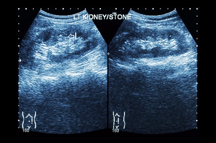
- Imaging tests: Screening tools such as abdominal X-rays, ultrasound scans, MRI (magnetic resonance imaging) scan or CT (computerised tomography) scans can assist with visualising the inside of the body and providing a doctor with insights into how the body is functioning and if there are any abnormalities causing symptoms. An X-ray may miss very small kidney stones, which can, however, be picked up in screening procedures like a CT scan. An intravenous urography or pyelogram / IVU or IVP (wherein contrast dye is injected into an arm vein to highlight the kidneys and entire urinary tract before X-ray visuals are taken) is another test which may be recommended. This modality is not the best at visualising a stone but is more accurate in showing one if there is swelling above the obstruction caused by a stone (this swelling is called hydronephrosis). Imaging tests, such as CT scans, can determine the presence of a kidney stone, where it is located and also determine its size. Signs of infection can also be determined using these tests. If there are large stones and signs of infection present, it may not be deemed safe to try and pass the stone naturally. Imaging tests can also be used to rule out other potential problems but can be limiting in terms of details seen if no contrast material (dye) is used.

