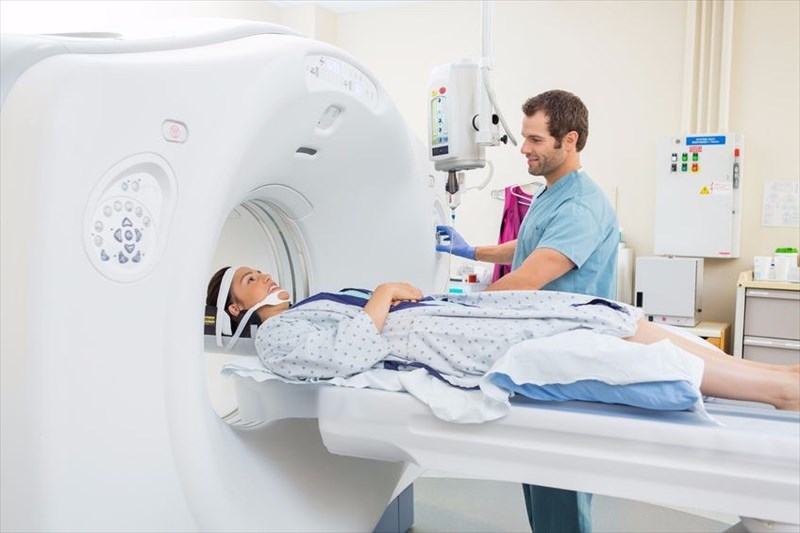
What happens during a CT scan?
A narrow X-ray beam is used to encircle the area of the body being analysed or assessed. During a scan, the beam will be used to provide a series of images, taken at different angles, that are then sent back to a computer. Technological software will then process the visuals and be sliced together to create a cross-sectional image of the inside of the body.
The process is repeated until enough images are produced to stack the scans, one on top of the other, forming the final 3-dimensional image (3-D visual). This visual essentially re-creates a 3-dimensional visual of your insides, such as an organ, set of bone structures or blood vessels. The visual can then be looked at from every angle to assess the nature of a medical problem or injury or diagnose a condition (if there is an abnormality present).
Often, a doctor will use a dye, known as contrast material (iodine or barium sulphate), just before a scan. This helps to highlight various areas of the body, such as tissues and easily shows up portions a doctor would like to assess more clearly. The contrast material appears white on the images produced and blocks X-rays, helping to provide emphasis where it is needed, and better enabling your doctor to better see what may be wrong.
You may be given contrast material ahead of your scan in one of three ways:
- Injection: An injection can be administered through a vein in your arm if your urinary tract, gallbladder, liver or blood vessels are to be scanned. During the injection, you may experience a warm sensation and a metallic taste in your mouth.
- Orally: A liquid containing contrast material, which is not typically pleasant to drink, may be given for you to swallow if your oesophagus or stomach is being scanned.
- Enema: If your intestines are to be scanned, contrast material may be inserted in your rectum. You may experience bloating in the abdomen, which can be a little uncomfortable.
Once a scan has been completed, you will be given plenty of fluids to help your kidneys flush out the contrast material from your system (i.e. expel the substance from the body).
The duration of a scan can take anything from a few minutes up to about 30-minutes, depending on the nature of the scan and the machine used. Scans normally take place in a hospital or an outpatient facility (such as a radiology clinic).
If you are anxious at any point during the build-up to the scan or while inside the scanner, you doctor may administer a mild sedative to help you relax. Your technician may try and talk to you initially to help you feel calm.
What does the scanner look like and what will I be required to do?
The scanner is shaped like a large doughnut type of tunnel with the hole in the centre. A narrow, motorised table slides through this hole or opening in the centre. You will be required to lie down flat on this table. The table has straps and pillows which may be used to assist with keeping you in the best position for the test, and a cradle that will help to keep your head still during the scanning process.
Once you are laying down flat on your back (facing upwards) and settled into position, the table will be moved slowly into the doughnut shaped scanner. An X-ray tube and detectors will then be activated to rotate around you (you will not be moving at all). The rotation action is what is capturing images and will make clicking, buzzing and whirring sounds during the scanning process. Each rotation will create thin slices of visuals for the creation of cross-sectional images to be stacked together.

You will be asked to keep as still as possible throughout the scan. Any movement can disrupt the visuals created and cause blurring. Each image taken needs to be as clear and detailed as possible in order to be accurately assessed. From time to time your technologist may request that you hold our breath during the scan. This is not unusual and you need not panic. It may be that visuals need to be taken of a specific area and the rise and fall of your chest as you breathe may cause blurring. You will only need to hold your breath for a few seconds at a time. The table may move a few millimetres at a time during the process. You will remain perfectly still the entire time.
A scan can take place within a matter of minutes and up to half an hour, depending on the nature of your scan and what is needed for assessment. The entire process is painless and non-invasive. You will not be required to stay at a hospital facility.
Once the scan has been completed, you will be able to resume your normal day’s activity. Before departing, you will be advised about how long it will be before you can expect results. If you were given contrast material ahead of your scan, you will be advised to linger a little longer and given plenty of liquids to drink to help expel the material from your body. Once medical staff are satisfied that you are well enough and haven’t experienced any adverse reactions, you will be allowed to leave the hospital or radiology clinic. You may be asked to drink more fluids through the day or night to help your kidneys flush out all of the contrast material.
If you require medical care following your scan or you have already been admitted to hospital for any reason, you will be taken back to your ward (room) for monitoring. In most cases, where you are merely having a scan done, you will be allowed home shortly afterwards.
If a young child is being scanned, he or she will be sedated and positioned on the table for the test. A little one will ‘sleep through’ the entire scanning process and be taken to a recovery area afterwards to rest peacefully while the sedative wears off. Contrast material will be flushed out soon after the scan and if well enough, a young child will be allowed home.
Are there any risks involved?
Risk is usually minimal but your doctor or the technician conducting the scan will explain all potential factors ahead of the scanning procedure.
These discussion points may include:
- Ionising radiation exposure: During the scanning procedure, you will be exposed to a low dose of ionising radiation. As the scan is designed to pick up detailed visuals, this amount of radiation is greater than that of a normal X-ray. The amount of exposure, however, has not been seen to cause any long-term effect. Risk may increase if you have multiple scans during your lifetime. Any increased cancer risk and damage to your DNA due to the exposure is very small and the benefits of the scan outweigh the low amount of risk potential. Risk may be higher for young children as they are still growing, but machines and dosages of radiation are usually adjusted for children to minimise potential risks. A general rule of thumb is that the higher the number of portions of the body being examined, the higher the radiation dose and thus, the higher the potential risk. Newer machines in use require less scanning time, which in turn means less radiation exposure.
- Pregnancy and an unborn baby: In most cases, a CT scan during pregnancy poses a very low risk of harm to an unborn baby. You should, however, advise your doctor of your pregnancy (should you not yet be showing). A doctor may opt for an alternative means of image testing, such as an ultrasound or MRI, if he or she feels it necessary to avoid radiation exposure altogether.
- Adverse reactions to contrast material: It can happen that you experience an allergic reaction to the contrast material administered before a scan. Typically, an allergic reaction is mild and merely shows up as skin itchiness or a rash. Rarely is an adverse reaction more severe, or even life-threatening. If you’ve experienced contrast material (or reacted badly to iodine) before and had a bad reaction, notify your doctor ahead of time to ensure your own safety during the test. Sometimes a doctor may recommend allergy medication or steroids as a way to counteract potential adverse reactions.
What other things may lead to adverse side-effects?
If you are diabetic and take medications as part of your treatment plan, you should notify your doctor. It may be necessary for you to stop taking your medication just before the scan and temporarily for a short period following the test in order to avoid an interaction between your diabetes medication and the iodinated contrast used that could cause lactic acidosis (a build-up of lactate in the body). Your doctor will advise when it should be safe to resume taking your medication.
It rarely happens that contrast material affects the kidneys and causes problems. If you’ve had kidney problems in the past, you should advise your doctor well ahead of time before the test.

